
Powering Cancer Progress
Meet the U of A researchers making strides in the detection, treatment and prevention of cancer.
Cancer is among the leading causes of death worldwide, claiming nearly 10 million lives each year, so it’s clear why it is a focus for scientists and researchers. But cancer also interests scientists because it is caused by the body’s own systems going haywire. Researching cancer is therefore a powerful tool both for understanding the human body and for creating real impact for millions of patients and families.
At the University of Arizona, researchers are diving deep into all types of cancer research. The university’s National Cancer Institute- designated Comprehensive Cancer Center, founded nearly 50 years ago, houses hundreds of doctors and researchers and has more than 70 labs participating in over 200 clinical trials. And there is even more cancer research happening at other campus institutions, like the Center for Advanced Molecular and Immunological Therapies (CAMI). Arizona researchers are making important discoveries that can help doctors be more targeted in their approach to treating cancers that affect specific groups of people. They’re also developing new technologies for diagnosing and preventing cancer.
WHAT A CANCER TELLS US ABOUT MEN
Dan Theodorescu, M.D., Ph.D., the Nancy C. and Craig M. Berge endowed chair for the director of the Cancer Center and a professor in the College of Medicine – Tucson,
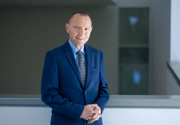
Dan Theodorescu
Photo: Joshua Elz
has been studying bladder cancer for decades. He has developed therapeutic techniques, discovered the genes that predict bladder cancer and developed
a personalized treatment called the Molecular Twin, an artificial intelligence- based precision oncology platform.
His recent study takes a new look at a well-known phenomenon that could expand our knowledge not just about cancer but about male aging in general.
“I’m pursuing a major discovery we reported two years ago, which showed for the first time that the Y chromosome is very important not only in cancer biology but also for therapeutics,” Theodorescu says.
The Y chromosome is significantly smaller than the X chromosome and does not encode nearly as many genes as X does. But now, Theodorescu’s cancer research is shedding new light on the importance of this chromosome.
“Loss of Y” is a condition where a man’s cells lose the Y chromosome. According to Theodorescu, it’s been known for a while that loss of the Y chromosome in certain tumor cells makes a person more likely to die of cancer.
The mechanisms behind why this occurs were unknown prior to Theodorescu’s findings in 2023 and 2025, which were both published in Nature.
“It turns out that the cancer that has lost the Y chromosome is more aggressive, because the immune cells that try to attack it become more exhausted and are having trouble controlling the cancer,” he says. “But there’s a silver lining.”
Cancer tumors with loss of Y are more sensitive to a common cancer immunotherapy treatment called immune checkpoint inhibitors. These inhibitors reverse the exhaustion of the immune cells and allow the body’s immune system to fight the cancer. Theodorescu’s new study found that the loss of Y in immune cells in the blood makes the immune system less effective at infiltrating the cancer.
A new treatment that has gained traction with cancer patients over the past few years that could be impacted by Theodorescu’s findings is called CAR T-cell therapy. This is a personalized cancer treatment in which a patient’s own T cells are modified in a lab to be better at fighting the cancer. Theodorescu discovered that if the cells modified by the lab are missing Y chromosomes, they are much less effective at fighting the cancer.
“The immediate implication is that people should be really mindful of what cells are used to generate CAR T cells,” he says. “Because you don’t want to have a CAR T product out of Y-negative cells.” Theodorescu is actively developing new therapies aimed at destroying cells that have lost the Y chromosome and also is looking into what causes loss of Y as men age.
GOING TINY TO STOP OVARIAN CANCER
Interim Vice Provost for Health Programs and Professor of Biomedical Engineering Jennifer Barton, Ph.D., has created the world’s smallest endoscope that can image and collect cells.
The equipment, less than one millimeter wide, is small enough to fit into a woman’s fallopian tubes and travel to the ovaries to look for ovarian cancer.
“Still today, there is no early detection method for ovarian cancer,” says Barton, who also holds the Thomas R. Brown Distinguished Chair in Engineering. “Symptoms are vague and usually aren’t diagnosed until the cancer is late stage.”
Because of this, many women with a family history of ovarian cancer have the ovaries and fallopian tubes removed at around age 35. Barton’s endoscope could help by identifying patients with abnormal cells early, eliminating the need for surgery on patients who don’t have abnormal cells.
Beyond the obvious challenge of making an endoscope smaller than the width of a credit card, the fallopian tubes also offer unique challenges.
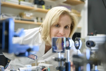
Jennifer Barton
Photo: Kris Hanning
“The tubes are very stretchy, and they’re not straight,” she says. “They can twist around and can change diameter. The fallopian tubes are also filled with all these finger-like projections that make getting through the fallopian tube more difficult than other small tubes of the body.”
Barton focused on overcoming the mechanical challenges, while waiting for laser fiber bundles to become small yet powerful enough for medical use, which took about 20 years.
Her endoscope is currently being used in two clinical trials, one in healthy patients to see how the endoscope works in the body and the other in fallopian tubes that have been removed from patients with ovarian cancer to confirm that they can visualize and collect the cancer cells.
She is working with Tech Launch Arizona to help find a manufacturer who can commercialize the equipment to get it in the hands of doctors within five years.
“The next step really will be to find a company that’s capable of producing this in quantity,” she says. “That’s just not something a university can do. I’ve got my graduate students down in the basement making them by hand.”
A VACCINE FOR CANCER
When most people hear mRNA, they immediately think of the COVID-19 vaccine. But mRNA has become one of the most powerful tools in a researcher’s tool belt. Tanvir Ahmed, Ph.D., for example, is using mRNA
to create medicine that is personalized to a patient’s specific cancer. His work at CAMI is creating a vaccine that trains the body’s immune system to target and destroy tumors.
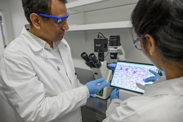
Tanvir Ahmed
Photo: Kris Hanning
“We are trying some unique technologies and platforms to figure out how we can successfully and effectively kill these tumor cells,” Ahmed says.
Ahmed explains that one of the reasons tumors grow uncontrollably is because they evade the immune system with a mutation. Traditional cancer treatments attack cancer cells, but they also attack healthy cells — they just deteriorate cancer cells faster because of their unregulated growth.
His mRNA cancer vaccine starts by collecting both healthy and cancerous cells from the patient and integrating the mutations from the cancer cells into the immune cells so the immune cells can then detect, target and destroy the cancer cells. It takes a few months to manufacture the personalized vaccine.
His team is focusing on melanoma because it is a very high intensity cancer, but he hopes to later develop vaccines for breast, prostate and colon cancer.
“If we develop this technology, it can target anything,” he says. He hopes to be in the first phase of clinical trials in three years.
A CANCER SCREENING WITHOUT THE DOCTOR’S OFFICE
Rogelio Robles-Morales, M.D., is a gynecologic oncology surgeon turned researcher. At the doctoral program in clinical translational sciences at the U of A School of Health Professions, he found a way to help cancer patients outside of the doctor’s office and far from the operating room.
He wanted to increase cervical cancer screenings and vaccinations in a community he is close to after growing up in Nogales, Mexico — Hispanic women.
According to Purnima Madhivanan, Ph.D., associate professor of health promotion sciences at the U of A’s Mel and Enid Zuckerman College of Public Health and a Cancer Center member, people from many cultures are not comfortable with pelvic examinations. Avoiding these invasive examinations keeps some people from identifying cervical cancer early and getting the health care they need.
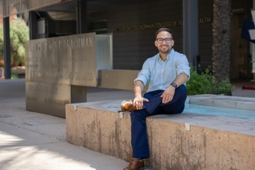
Rogelio Robles-Morales
Photo: Quion Lowe
“Only about 5% to 10% of women are at high risk for cervical cancer,” Robles-Morales says. “But you make all of them go through an invasive procedure that requires a clinical setting that has a high mental burden, instead of just bringing those who are positive on a screening.”
Robles-Morales wanted to create a cervical cancer screening that was less invasive, more comfortable and more likely to be usable for underserved communities.
He is testing three less-invasive field kits for collecting samples for cervical cancer testing: a vaginal self-sampling kit called the Simpli-Kit, a noninvasive urine collection and preservation kit called the Colli-Pee, and the Tasso, a self-collected blood sample kit.
His team collects and delivers samples from participants, then conducts in-depth interviews to determine which method was preferred, whether samples were collected correctly and which method participants were most willing to use.
They also look at whether the kits yielded accurate results. “I look for strategies that were high on recruitment,” he says. “Which one would the community gladly use, because we still have a problem with uptake.” ❖
We are grateful to the many Cancer Center donors whose generosity advances research in cancer prevention, detection, and treatment. Follow the QR code to learn more about exciting work at the Ginny L. Clements Breast Cancer Research Institute and to make a gift to support it.


