
Optics Solutions for High-Tech Health Care
BIO5 Institute researchers are making tools better and more accessible.
The BIO5 Institute is the University of Arizona’s home for cutting-edge research, often leading to innovative technology that is changing how science is done and ultimately impacting the way we live and work.
The institute, funded by Arizona’s Technology and Research Initiative Fund (TRIF), is designed to be the university’s cross-disciplinary hub for the biosciences and to be of service to our state.
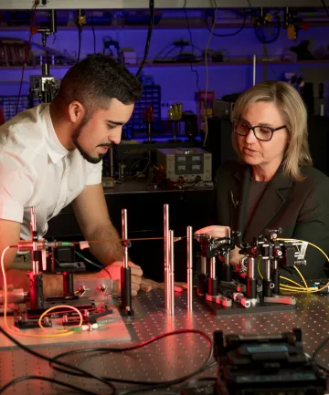
Jennifer Barton with Ricky Cordova, a biomedical engineering research specialist
BIO5 connects and mobilizes hundreds of world-class researchers — including bioscientists specializing in plants, animals and humans as well as engineers, physicians and computational researchers — to develop creative solutions for complex challenges such as disease, hunger, water and food safety, and other health issues facing Arizona and the world. This interdisciplinary approach has resulted in disease prevention strategies and promising new therapies, innovative diagnostics and devices, and improved food crops.
“We are able to help connect, facilitate and deploy people, resources and funding to produce a result that could help change the course of our community impact related to grand challenges like COVID-19 and other large-scale challenges and crises,” says Jennifer Barton, director of the BIO5 Institute.
In 2019, BIO5 opened its new 150,000-square-foot, $107 million Bioscience Research Laboratories to house about 50 principal investigators and nearly 400 researchers, including about seven graduate students per lab.
The new quarters connect with the 180,000-square-foot Thomas W. Keating Bioresearch Building, which opened in 2006 and now houses some 40 PIs and labs.
BIO5’s director, Jennifer Barton, serves as the chief connector of nearly 350 top scientists from over 70 departments across the university. Their collective bioscience breakthroughs have made possible new technology that is already making a mark in global markets. Barton, who has directed the institute since 2015, works on early cancer detection in her own BIO5 lab.
As a child in Fullerton, California, Barton spotted her first laser during a lesson in a sixth grade classroom at Golden Hills Elementary School. “I went to the library and looked them up in National Geographic magazine,” she recalls. Then she saw a laser light show as part of the U.S. Bicentennial celebration on July 4, 1976. “I was fascinated. They bounced lasers off the Washington Monument, and it was beautiful. That was what got me interested in working with lasers.”
Years later, she decided to study electrical engineering, getting a master’s at the University of California, Irvine, while working at McDonnell Douglas. She did a master’s report at UCI’s Beckman Laser Institute about lasers in medical technology, specifically dentistry. And while at McDonnell Douglas, Barton worked for six years with NASA on electrical power systems for the International Space Station.
In 1994, she started her Ph.D. in medical optics, working with A.J. Welch, “one of the grandfathers of lasers in medicine,” she says, in his lab at the University of Texas at Austin. Then she was recruited to the University of Arizona to help start a biomedical engineering program, which grew into a full department.
We asked Barton to take us on a conceptual tour of some of BIO5’s optics labs.
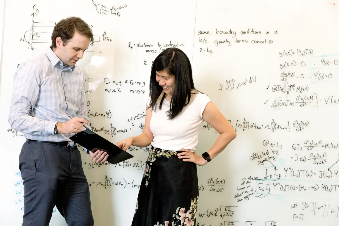
Euan McLeod, assistant professor of optical sciences, and Judith Su
Medical Imaging and Microfabrication
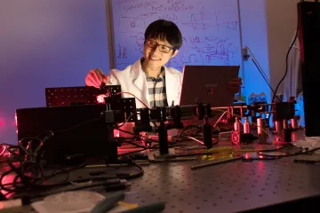
The tour began with the lab of Tsu-Te Su, known as Judith, who works on the microfabrication of nanostructures. “She makes incredibly sensitive detectors of biomarkers in the body. One of her big areas is looking at markers of Alzheimer’s disease.”
Su’s team is putting photonics technology into a biosensor that could become a useful diagnostic tool. It uses microtoroids, tiny ring-shaped structures, with which a light can be directed to travel around a sample and find very small particles of the protein biomarker being flowed past the sensor.
“She needs to fabricate these devices, and then we need to test them to make sure they are useful as a diagnostic,” Barton says. “Judith works with the physicians and brain scientists here in the BIO5 Institute to make sure her biophotonic device works for this medical problem.”
Optomechanics
The work of Dongkyun Kang, known as D.K., is an ideal example of where optics is going, Barton says. “D.K. is taking the confocal microscope and turning it into a portable handheld device that can go into rural areas — including rural Arizona and into developing countries — to diagnose cancers that affect a lot of people.”
Dongkyun Kang
Kang set out to do human tissue imaging in vivo using a smartphone attached to a confocal microscope. That could facilitate easy-to-use, high-resolution imaging in rural clinics at low cost — and ultimately allow for early diagnosis and treatment and lower mortality rates.
He calls it “stopping cancer with a smartphone.” The cancers Kang is targeting include skin cancers, cervical cancers and oral cancers.
“This shows what we are going to do in optomechanics,” Barton says.
The commercial confocal microscope is a large device that can cost upwards of $100,000. Kang is taking that capability and making sensors that are portable and highly sensitive — with a cost goal of $5,000.
“This is all because of advances in photonics,” Barton says. “You can use a small laser diode and implement it in the imaging and diagnostic devices that we are building. Today you can manufacture small optics at a reasonable price.”
Barton predicts an explosion of new technologies coming on the market. “I think we are finally there in terms of products that are truly useful, can truly help patients, and are inexpensive enough to have good market penetration. That’s the difference between the $100,000 device for confocal microscopy and the $5,000 device.”
Translational Imaging
In Barton’s own lab, work on spotting ovarian tumors is unfolding. She is developing what will be the first effective screening method for ovarian cancer. The work, she says, illustrates BIO5’s ability to carry out fabrication and imaging using high resolution optical techniques.
“If you want to look at cells or tissue microstructure, you can’t image very deeply. You end up needing to build endoscopes,” Barton says. “And I do that.”
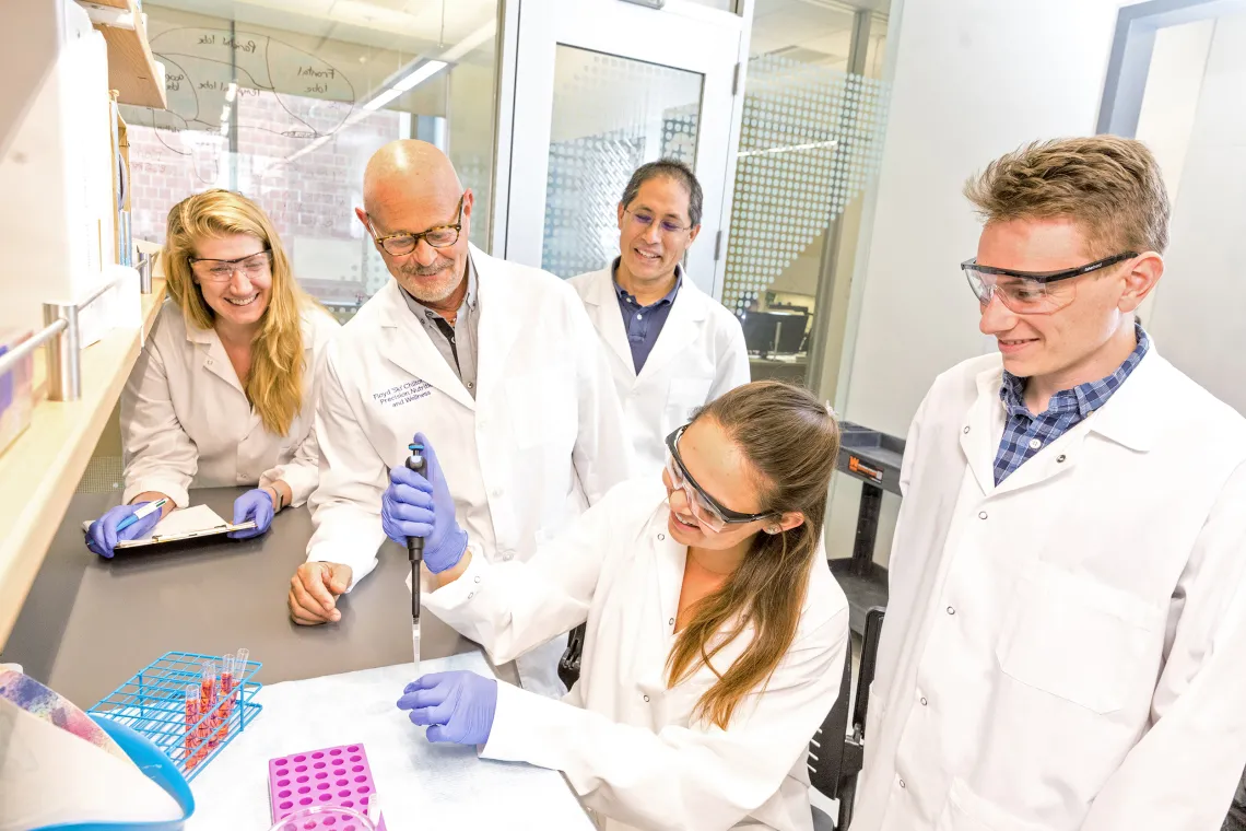
Researchers at BIO5
Her microendoscopes provide nonsurgical techniques for looking into the body’s tissues.
The body has many natural orifices needing study, Barton says, “and the same advances in photonics and materials allow us to make tiny, flexible endoscopes that can go all kinds of places in a body, in a pretty minimally invasive manner.”
Barton’s work, funded by a Department of Defense Ovarian Cancer Research Program grant, has involved placing the devices in the fallopian tubes, where they can do imaging below the surface to a depth of about a millimeter with noninvasive optical coherence tomography, or OCT.
Previous technology only allowed the taking of white light images on the surface. “We are now going to create images like what your eye would see if it could get in there,” Barton says.
The new technology, which Barton says is about seven years away from becoming a market product, will provide the capability to look for cancer below a surface with OCT, to look at cell functions and metabolism with fluorescence, and to look at cell structure with microscopy. “We are adding a great deal of extra capability to what the medical community has had in the past.”
Tissue Optics
Because skin cancer is widespread in Southern Arizona, BIO5 has a strong skin cancer imaging program. Its labs are looking at how to use tissue optics to diagnose skin cancer, which currently often involves taking a biopsy.
“How can you diagnose skin cancer without having to take a biopsy?” Barton says. “Melanoma, of course, is the killer, but how do we figure out the hallmarks of whether tissue is benign versus a melanoma?”
BIO5 also studies how to treat cancers with photodynamic therapy. Work done by Clara Curiel, a physician and dermatology professor, centers on diagnosis and treatment for squamous cells and basal cells, which represent a huge public health cost burden in Arizona and Australia.
What links the work of hundreds of researchers at BIO5 is a shared focus on bringing technology out of the lab and into the field. “We’ve had plenty of time for optical technologies to develop lots of capabilities,” Barton says. “And the capabilities are great: We are there; we can image with high resolution and high sensitivity. Now we need to focus on making them more accessible. And that means it’s got to be less invasive and less expensive.
“To me, that’s what we should be doing in the 2020s.”


